Cell biology
Animal cells
A closer look at the small, busy structures that make up animal life.
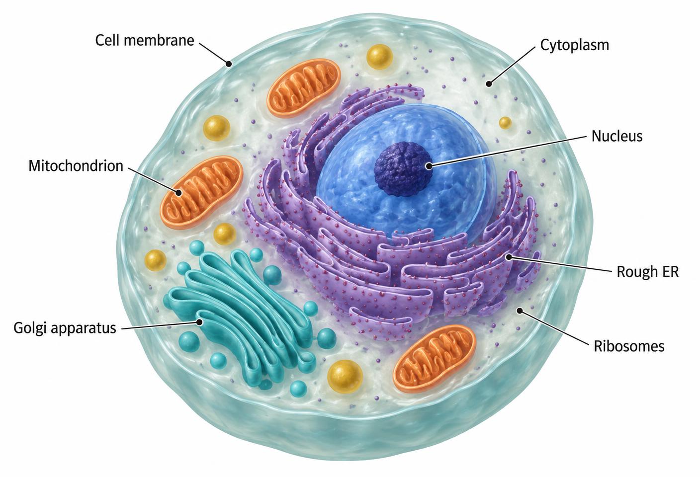
Inside a living cell
This cutaway opens up an animal cell so its inner structures can be seen together. The cell membrane forms a flexible boundary that controls what enters and leaves. Inside it, the cytoplasm surrounds the organelles, and many chemical reactions take place in its fluid portion, the cytosol. The colours help distinguish the structures; real cells are not coloured like this, and their shapes vary with their roles.
The large blue structure is the nucleus, which contains most of the cell's DNA. The darker spot inside it is the nucleolus, where parts of ribosomes are assembled. Ribosomes build proteins by joining amino acids together. Some float freely in the cytosol, while others sit on the folded membranes of the rough endoplasmic reticulum, often shortened to rough ER.
The curved turquoise stacks are the Golgi apparatus. Proteins made through the rough ER can travel here in small membrane-bound sacs called vesicles, then be modified and sorted for their next destination. The orange mitochondria supply much of the ATP that powers cellular work. Each organelle has a different role, but the cell depends on them working together rather than acting as separate machines.
The bigger picture
A muscle cell, a nerve cell and a skin cell share much of this basic organisation, yet differ in shape and in the amounts of particular organelles. Their structure reflects the work they do. Unlike plant cells, animal cells do not have chloroplasts or a cellulose cell wall.
Key terms
- Organelle
- A specialised structure within a cell.
- Nucleus
- The compartment containing most of an animal cell's DNA.
- Ribosome
- The structure where proteins are assembled.
- Vesicle
- A small membrane-bound sac that stores or transports material.